

Since our beginnings, at DrAW Clinic we have worked to be pioneers in the adoption of new technologies.
These, on the one hand, have allowed us to offer greater comfort and safety to our patients and, on the other hand, have helped us to be more accurate in diagnosis and more effective in treatment.
As a sign of this firm commitment to offer patients the latest advances in the field of dentistry, we have recently acquired new equipment.
Among them, we would like to highlight two 3D intraoral scanners for Orthodontics, a 3D intraoral scanner for Implantology and a microscope for endodontics.
Technology in Orthodontics
All orthodontic treatment requires a rigorous preliminary study so that the specialist can plan it in the best possible way.
In this way, optimal results can be obtained according to the requirements of each person.
To this end, and also in order to facilitate the visit to patients, we have two specific scanners for this specialty.
3D intraoral scanner
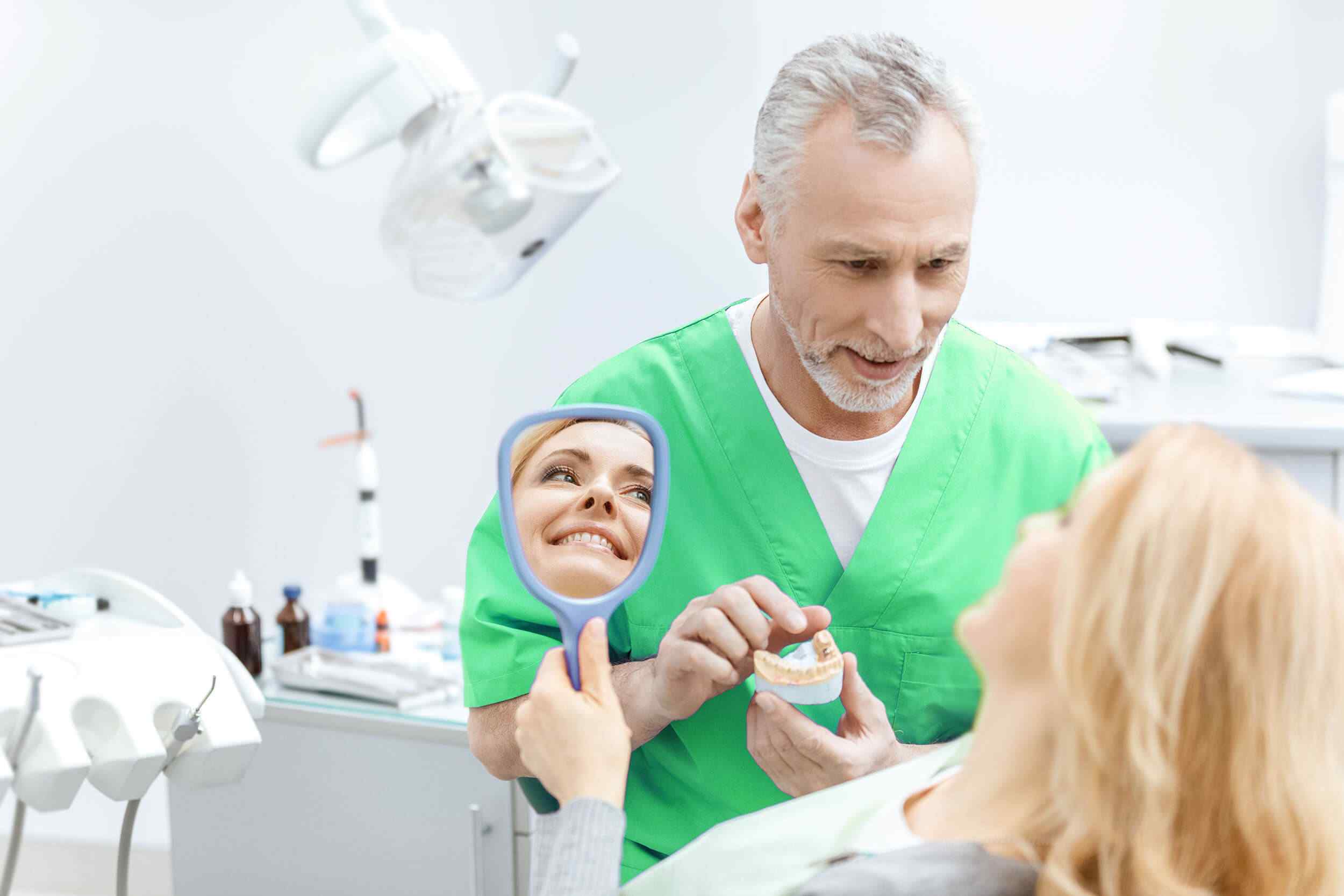
The 3D intraoral scanner used by our orthodontic team is the one that has been in use the longest at DrAW Clinic.
After several years working with this instrument, there is no doubt about its numerous benefits.
This device allows us to obtain 3D digital impressions of the mouth of a patient who is going to wear orthodontics with lingual brackets -Incognito- or with Invisalign aligners.
The scanner has a pencil that we pass over the different faces of the tooth to obtain a recreation of the mouth in real time.
As we move forward, this image appears on the computer screen and, with this, our orthodontists can see both the imperfections that the patient has in the enamel and the malpositions.
Once we have passed the pencil over all the faces of the teeth, the scanner creates a model from which we can order both Incognito brackets and Invisalign splints from both brands.
The technology used makes this a very accurate method, as well as very quick to start treatment.
This is because the brands -located outside Spain- receive the digital image immediately.
Thus, the dental appliance – splints or lingual brackets – can begin to be manufactured much more quickly, since the models do not have to be sent physically.
You will be interested in ” Piezosurgery in maxillofacial surgery: what are its advantages?
On the other hand, the use of the scanner is very convenient for the patient, since we avoid having to make the old impressions.
The most traditional technique consisted of introducing a mold with alginate paste in the mouth, which sometimes generated a choking or nausea sensation.
Intraoral 3D scanners and microscopes bring extra value to the treatment, facilitating diagnosis and interventions.
Find out what your smile will look like with iTero
One of our most recent additions is the iTero Element scanner, whose main feature is very attractive to patients.
It allows you to get a simulation of what your mouth will look like after orthodontic treatment, even before you start it.
The iTero scanner has a constant flow of digital information from a gigantic database of Invisalign, the brand that markets this equipment.
Thanks to artificial intelligence and continuous connection, it searches and compares cases similar to the person’s to show a color simulation of the result.
Oral simulation with iTero
Enlarge image
ITERO INTRAORAL SCANNER
3D intraoral scanner for implant dentistry
Like the one we use in Orthodontics, what the Implantology scanner does is to show a three-dimensional recreation of the patient’s mouth.
However, it has the particularity that it is in color.
This is a detail that is not important for orthodontic treatment, but it is fundamental for a person who is going to undergo implant surgery.
With this scanner, it is possible to capture the real colors of the teeth so that, subsequently, we can manufacture a crown that is the same shade and fulfills a highly esthetic function, as well as functional.
With the use of this scanner, we also achieve a more accurate 3D model than could be obtained with traditional impressions.
Thus, the fit of the crowns is improved by being more precise in capturing the margins.
In this way, we can prevent the crown from loosening or decementing later on.
In addition, we offer greater comfort to the patient, since we do not have to place the conventional molds with the paste.
Other advantages
As with the orthodontic scanner, it also makes the procedure for placing the final crown faster, since the transmission of the work is telematic.
Finally, another advantage for the patient is that the scanner records all the necessary information about the process (data of the crown, the time when it was made, measurements…).
This point is very important in the event that the crown has to be repeated or the patient changes residence, for example, by moving abroad.
The virtual models could be sent to another practice so that when the patient arrived, the new crown would already have been made.
Although scanners are a CAD-CAM technology that is highly developed in other industries, their use in the dental sector is relatively new.
In fact, the 3D intraoral scanner that we have acquired for Implantology is the latest model on the market and very few clinics in Spain have this technology.
Ask about the technique
As well as the skill of the professional, the fact of having advanced technology allows to obtain better results.
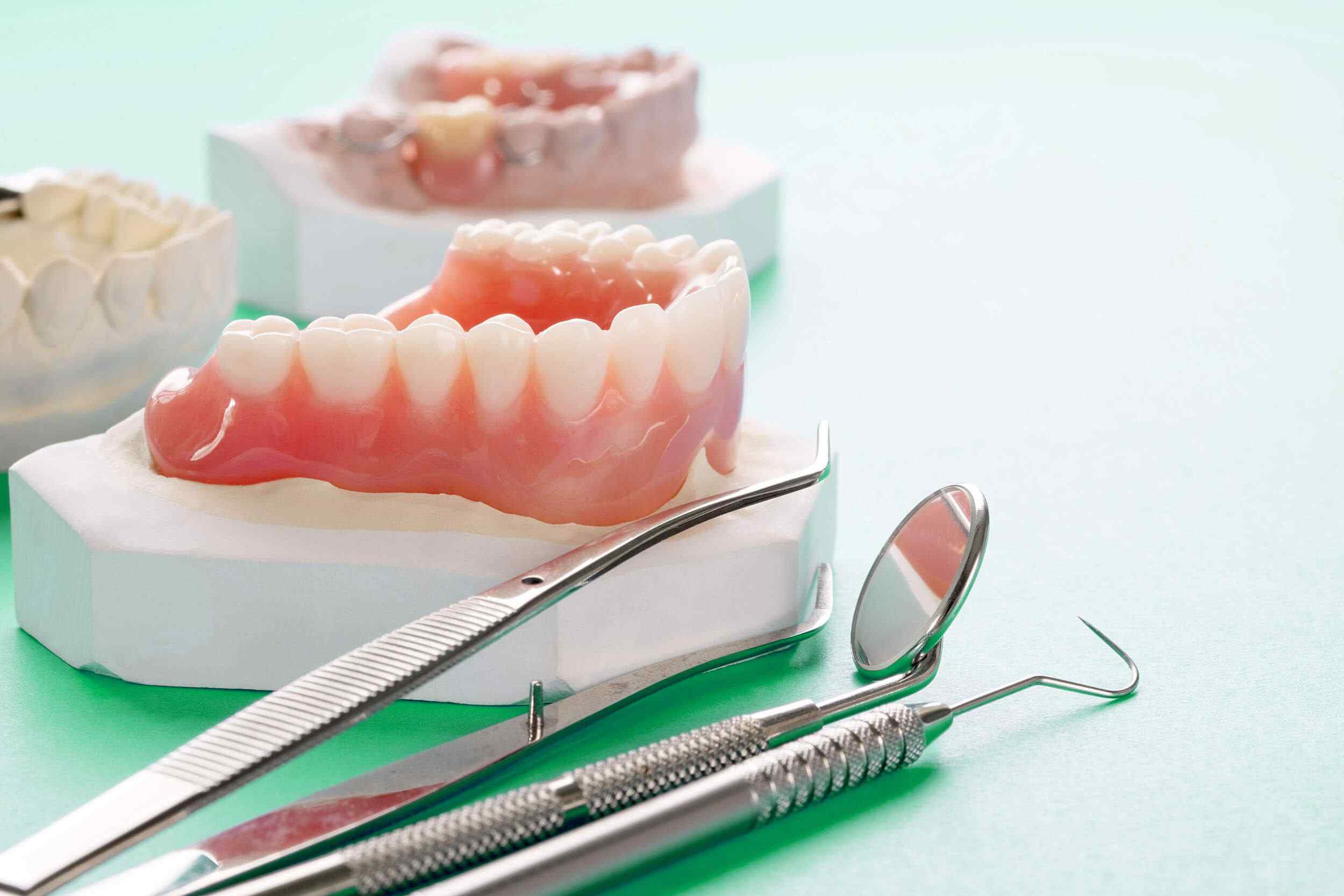
General Dentistry Microscope
The microscope we have at DrAW Dental Clinic is of Spanish manufacture and we use it to carry out both endodontic and periapical surgeries.
The main advantage it offers is the magnification it allows us to achieve, which is 250 times human vision.
This magnification clearly improves the predictability of treatment and ensures that we can carry out minimally invasive dentistry.
At this point, we must not forget that both endodontics and periapical surgery are complex treatments because dentists have to work in small, very limited and dark areas.
Without a microscope to provide greater vision and illumination of the area, the only help the professional has is his or her own “touch” and the X-rays taken of the patient.
Although this has been sufficient in many cases that have been successfully resolved, there is no doubt that the magnification obtained with the microscope contributes in a very positive way to increase the success rates and predictability of the treatments.